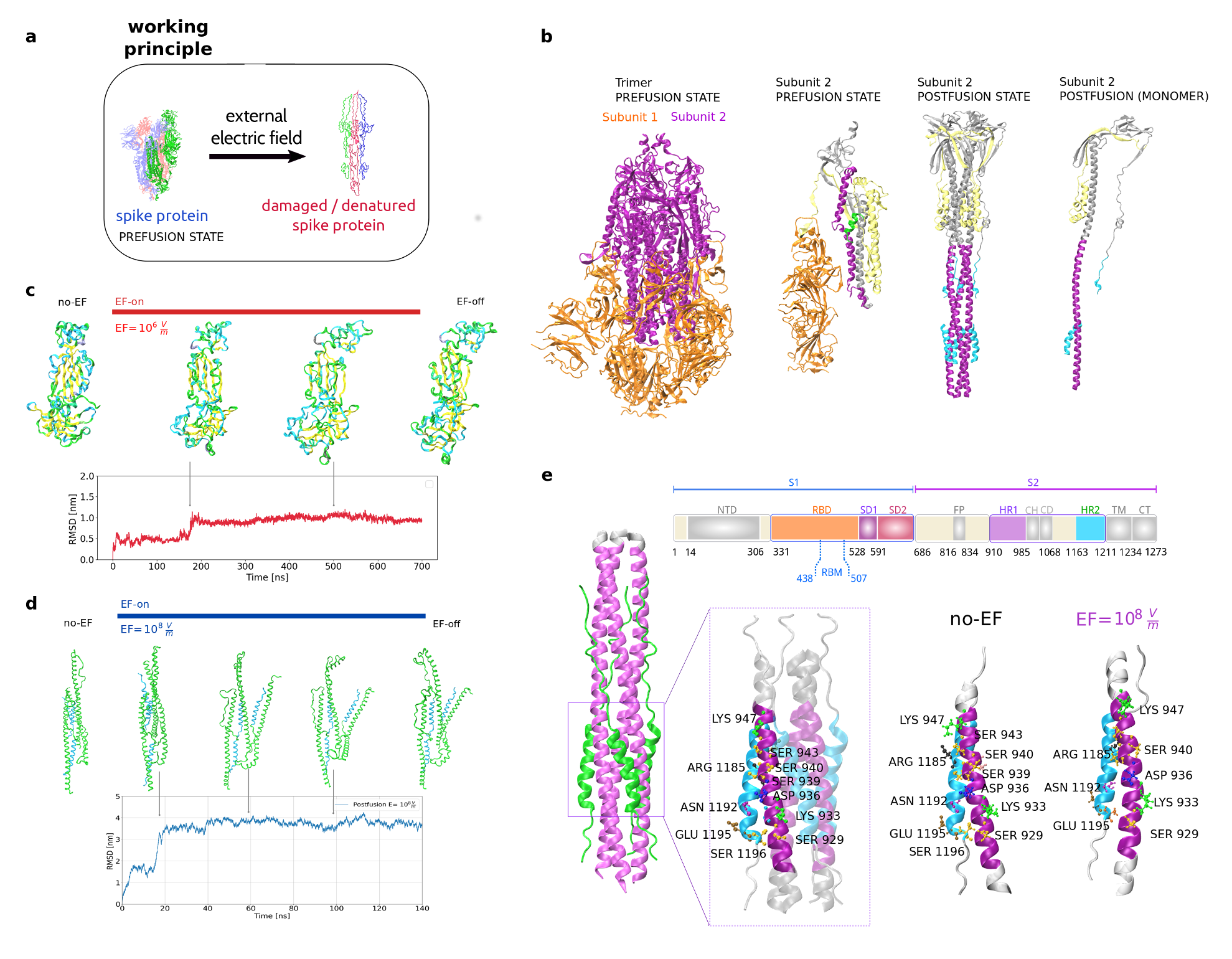
Figure 1:
a) The Spike protein in the prefusion state, present in the SARS-CoV-2 virus envelope, can be altered when external electric fields (EF) induce drastic conformational changes and damage to its structure.
b) Snapshots of the studied subunits of S: subunit 2 in prefusion state (PDB 6VSB) and subunit 2 in postfusion state (PDB 6LXT). The major shape changes occur in the different subunits and between subunits of the S protein in the prefusion state.
c) Snapshots of the prefusion conformation (S1 subunit) as it evolves with EF application (upper row). Deviations from the initial structure are quantified with the Root Mean Square Deviation (RMSD) (lower row).
d) Snapshots of the postfusion conformation (S2 subunit, PDB 7COT) under EF (upper row) and deviations from initial structure as RMSD (lower row). No changes in the secondary structure were observed throughout the MD trajectory of the postfusion structure, despite the presence of a high-intensity field.
e) Schematic representation of the S1 and S2 subunits within the SARS-CoV-2 Spike protein. The segments used in this study have been distinctly highlighted using colors (upper panel). The core of the S2 subunit in the postfusion state (PDB 6LXT) and structural details of the region of chains interactions are presented in cartoon form (PDB 7COT). Important residues are indicated and labeled. The fusion core regions for each protomer remain conserved after the application of EF.